There is a 12' Japanese Maple tree in our neighborhood.
Several years ago I noticed the bark was beginning to split along some branches
that were several inches thick.
Yet the foliage looked fine, and most people would have said it was a beautiful, healthy tree.
Last year the bark splitting was worse, and about 20% of the tree was dead.
Strangely, it retained many of its dead leaves over the winter, something it had never done before.
I figured that was not a good sign.
Sure enough, this spring (2008) the tree is now about 80% dead.
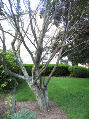
1
This picture was taken in mid-July, when the tree should have been fully leafed-out.
But as you can see, the tree is now mostly dead.
In particular, note the shedding of bark at the trunk of the tree.
A rubbing branch a few feet above this area probably made it easier for disease to enter the tree
several years earlier.
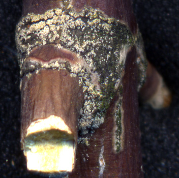
2
There is a heavy concentration of spores on the scars at the branch junctions,
as shown in this view. Strangely, spores are not present on the surrounding
bark, as on other trees.
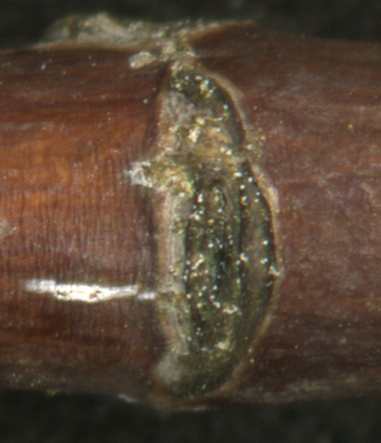
3
There were also spores at the leaf scars, as shown here.
The remainder of the branches had very few spores on them.
While pictures 2 and 3 were taken in late May, the following set of 400x twig
and leaf cross-section microscopic pictures
were taken in late September.
At this time the white canker had already reproduced by shedding it's spores.
Hence, spores were hard to find.
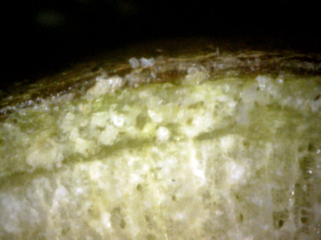
4
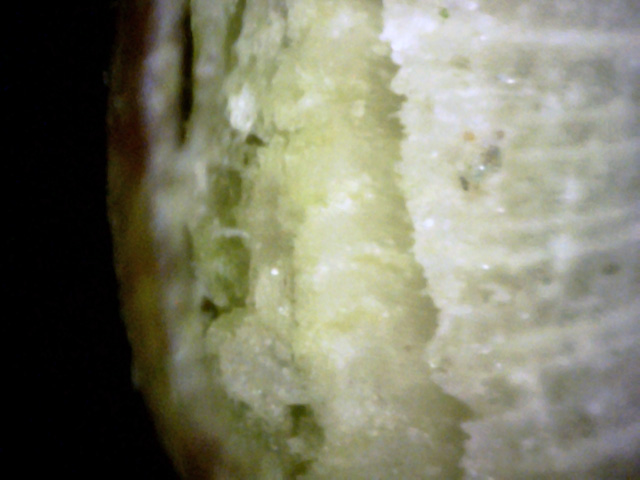
Here is a section of sapwood riddled with canker particles.
They are also present on the inner bark, and some can be seen on the outer bark.
 →
→
5
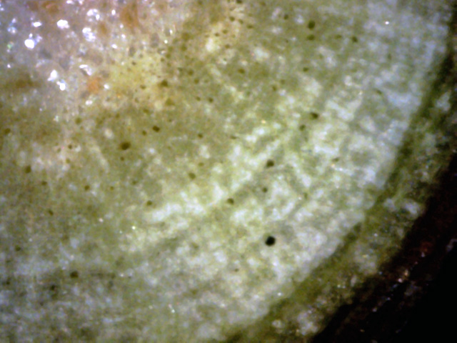
The inner bark has been greatly mangled by canker growth.
In particular, note the blob of gray canker material near the bottom of this view
(red arrow).
It has that typical foamy look.
6
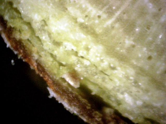
An unusually insightful view showing a number of growth rings, each infected with white canker.
It shows this twig was infected with white canker about three years ago,
which is about when the tree's decline started.
Also notice the patches of white canker infection in the outer bark.
 ←
←
7
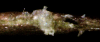
The item of interest here is that gray-white blob at the end of the void under the bark (red arrow).
Is it a canker eating the sapwood or an insect eating it?
8
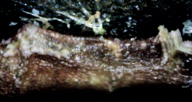
In this leaf cross-section, we see a typical example of a white canker growth extending
completely through the leaf, and the the canker on each side of the leaf is starting to bud off hypha.
9
The interesting part of this picture is that there is a large spore collection on the left
which is bulging out of the leaf, and it seems to be connected within the leaf to the
parallel set of white tubes emerging upward on the right.

10
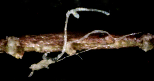
A hypha runs through the leaf from the upper-left to a canker blob at the lower right.
This blob is emitting a hypha from its tip.
The hypha in turn has turned upward to touch the leaf surface, and has begun to infect it with what looks like a spore.
11
Great example of two white canker blobs, each connected to a hypha.
The blob's granular appearance hints that it envelops the leaf cells, digesting them in order
to get energy to send out hyphae, which in turn spreads the infection.
This once beautiful Japanese Maple is now mostly dead.
The presence of white canker spores and white canker growth are evidence this tree is being killed by white canker.
This tree has been in steady decline for about three years, as have other Japanese Maple trees in the area.
The first visual disease clue was splitting bark.
Another visual clue was the death of random branches.
Yet another visual clue was the failure of the tree to shed its dead leaves during late fall.
The relatively rapid decline of this tree compared to other species lends weight to the belief
that white canker affects maples to a greater extent than other species.